
The research is devoted to the study of the causes of spinal pathology, including scoliosis, pain syndrome, and other conditions, as well as their relationship with disorders of the internal organs. Clinical and experimental exploratory studies led Prof. V.V. Serdyuk to the scientific discovery titled "The Pattern of Development of Diseases of the Internal Organs in Adults as a Consequence of Spinal Deformity Originating in Childhood." Diploma No. 1, issued by the Ukrainian International Academy of Original Ideas on December 1, 1995.
The second discovery in the field of vertebrology is devoted to Prof. V.V. Serdyuk's development of a new theory of the origin and progression of idiopathic scoliosis: "The Pattern of Development of Scoliotic Deformity of the Spine Based on Zones of Instability in All Its Regions, Arising as a Consequence of Unilateral Hypertonicity of the Back Muscles Associated with Asymmetric Function of the Cerebral Hemispheres." Diploma No. 5, issued by the Ukrainian International Academy of Original Ideas on January 16, 2008.
An analysis of Prof. V.V. Serdyuk's own clinical material, comprising more than 6,900 patients with idiopathic scoliosis and spinal pain syndrome, accumulated between 1996 and 2010, is presented in the monograph "Body Asymmetry. Scoliosis. Spinal Pain Syndrome. A New View of an Old Problem," Donetsk, 2010, 392 pages.






A detailed description of the essence of the new theory concerning the origin and development of idiopathic scoliosis is presented in the scientific article by V.V. Serdyuk, "Idiopathic Scoliosis. Mechanisms of Its Development," Kyiv, Bulletin of Orthopaedics, Traumatology and Prosthetics, No. 3, 2010, pp. 19-26. DOWNLOAD PDF
In addition to the above, since 1974 Professor V.V. Serdyuk has worked as a specialist in reconstructive orthopaedic surgery and has accumulated unique experience in the surgical treatment of disabled patients with flaccid paralysis of the lower limbs resulting from spinal cord injury or poliomyelitis. The Department of Orthopaedics and Traumatology, supervised by Prof. V.V. Serdyuk, is part of the Odesa Traumatology Center, directed by Professor Yu.V. Sukhin, and is one of the leading medical institutions in Ukraine where this type of treatment is performed.
Effective surgical procedures allow paralyzed disabled patients to begin walking independently with the aid of crutches and, subsequently, after restoration of muscle strength in the limbs, without them. After such treatment, former patients begin driving cars, acquire professions, including the medical profession, return to active social life, and become fully integrated members of society.
Prof. V.V. Serdyuk actively participated in the development of a complex of surgical procedures for the treatment of children and adults with disabilities suffering from the consequences of cerebral spastic paralysis.
Prof. V.V. Serdyuk and his colleagues successfully treat almost all types of post-traumatic, congenital, and acquired deformities, including flatfoot, hallux valgus deformity of the first toe (commonly referred to as a bunion), degeneration of the palmar aponeurosis with Dupuytren's contracture, flexion and extension contractures of the limbs, and other conditions. Lengthening of the upper and lower limbs is also performed.
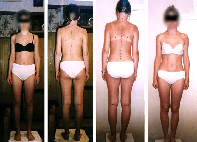
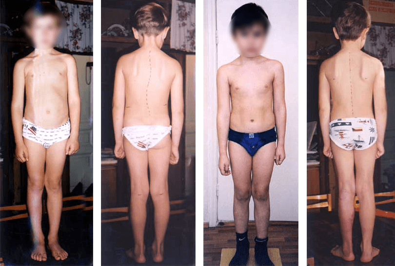
Complaints of headaches, emotional lability, manifestations of vegetative-vascular dystonia, and pain in the arms and legs. Relative shortening of the left leg by 37 mm. Initial condition shown in the two photographs on the left. Hypotrophy of the left gluteal region. Long-term result: the third and fourth photographs from the left, taken 1.5 years after the start of treatment. Complaints were absent, the spinal axis was correct, and leg length was equal. The volume of the right and left gluteal regions was practically identical.
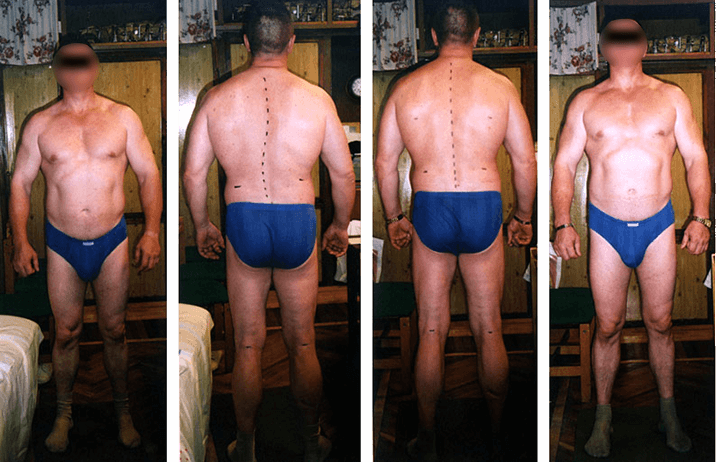
Periodic pain in all regions of the spine, as well as in the lower limbs after physical exertion. Relative shortening of the left leg by 18 mm. Initial condition shown in the two photographs on the left. Hypotrophy of the left gluteal region. Long-term result: the third and fourth photographs from the left, taken 6 months after the start of treatment. The spinal axis was correct, leg length was equal, and there were no complaints. Asymmetry of the gluteal muscles was practically absent.
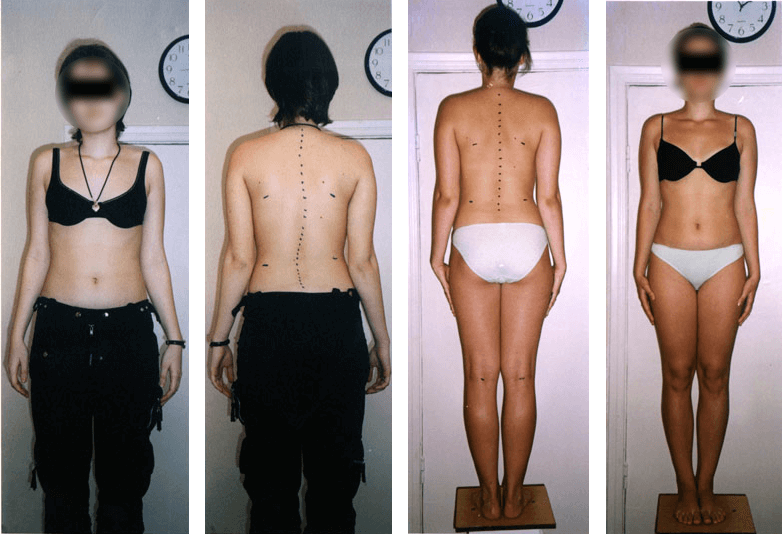
Torticollis, impaired vision, eye irritation, pain in the arms and legs, rapid fatigability, and vegetative-vascular dystonia. Relative shortening of the left leg by 10 mm. Initial condition shown in the two photographs on the left. Hypotrophy of the left gluteal region. Long-term result: the third and fourth photographs from the left, taken 6 months after the start of treatment. The spinal axis was correct, shortening of the left lower limb was absent, and complaints were absent. The volume of the gluteal muscles on the right and left was practically identical.
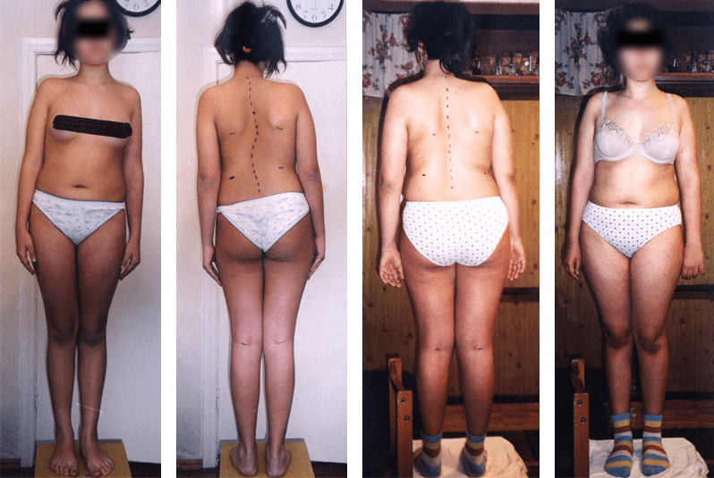
Relative shortening of the left leg by 40 mm. Persistent headaches, impaired vision, pain in all regions of the spine, as well as in the arms and legs. Initial condition shown in the two photographs on the left. Marked asymmetry of the gluteal regions and of the left and right halves of the pelvis. Long-term result: the third and fourth photographs from the left, taken 1.5 years after the start of treatment. The spinal deformity was eliminated. The general condition normalized, complaints were absent, and headaches were absent. Asymmetry of the left and right halves of the pelvis was mild.
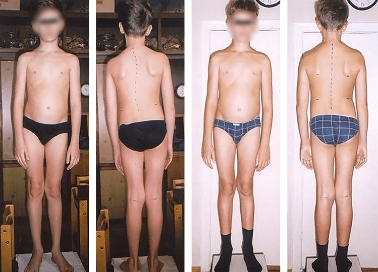
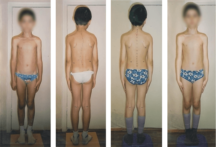
Impaired vision, convergent strabismus, vegetative-vascular dystonia, and persistent headaches. Complaints of leg pain, bilateral lumboischialgia. Two years before presentation, the patient had been treated for cholecystitis, duodenal ulcer, pyelonephritis, and cystitis. Relative shortening of the left leg by 15 mm. Initial condition shown in the two photographs on the left. Marked pelvic asymmetry with severe tilt to the left. Long-term result: the third and fourth photographs from the left, taken 1.5 years after the start of treatment. The spinal deformity was eliminated, and posture became correct. The position of the eyes normalized. Complaints were absent. Mild asymmetry of the gluteal muscles persisted despite correct pelvic position.
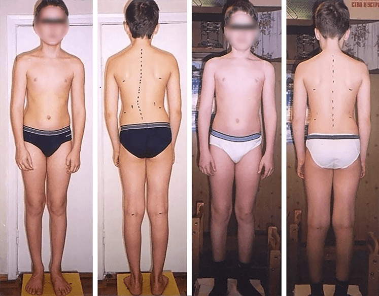
Impaired vision, convergent strabismus, vegetative-vascular dystonia, and headaches. Initial condition shown in the photograph on the left. Two months after completion of treatment, shown on the right. Result: the spinal deformity was eliminated. Posture became correct, the position of the eyes normalized, and complaints were absent.
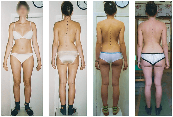
Severe spinal pain syndrome. Persistent headaches and rapid fatigability. Signs of neuritis of the left trigeminal nerve. Numbness of the hands and pain in the heart region. Bilateral plexitis and lumboischialgia. Relative shortening of the left leg by 30 mm. Initial condition shown in the two photographs on the left. Marked hypotrophy of the left gluteal region. Long-term result: the third photograph from the left, taken 1 year and 7 months after the start of treatment. The spinal deformity was eliminated. Posture became correct. Complaints were absent. The fourth photograph from the left, taken 2.5 years after the start of treatment. Complaints were completely absent. Leg length was equal. The volume of the right and left gluteal regions was restored.
Pain in the arms and legs. Rapid fatigability. Initial condition shown in the photograph on the left. One year after completion of treatment, shown on the right. Result: the spinal deformity was eliminated. Complaints were absent.
Amblyopia and astigmatism. Vegetative-vascular dystonia. Convergent strabismus. Pain in the arms and legs. Initial condition shown in the photograph on the left. Six months after completion of treatment, shown on the right. Result: the spinal deformity was eliminated. Vision normalized. No complaints were reported.
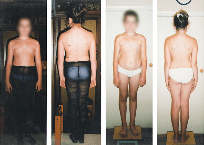
Biliary dyskinesia. Latent convergent strabismus. Vegetative-vascular dystonia. Pain in the arms and legs. Periodic pain in the heart region. Initial condition shown in the photograph on the left. Eight months after completion of treatment, shown on the right. Result: the spinal deformity was eliminated. Vision normalized. Complaints were absent.
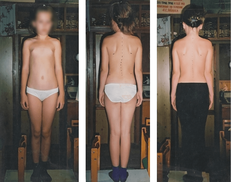
For two years, the patient stayed in a special boarding school for children with scoliosis, but no positive results were achieved. Pain in the left leg. Retrosternal pain in the heart region. Rapid fatigability. Initial condition shown in the photograph on the left. Six months after completion of treatment, shown on the right. Result: the spinal deformity was eliminated. The patient reported no complaints.
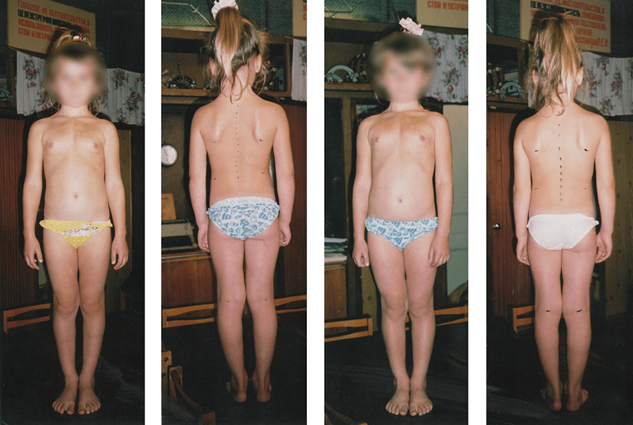
Vegetative-vascular dystonia. A cyst of the left ovary was detected, and the elevated estrogen level had been attributed to its presence. Pain in the legs. Initial condition shown in the photograph on the left. Four months after completion of treatment, shown on the right. Result: the spinal deformity was eliminated. Complaints were absent. Ultrasound examination confirmed resorption of the ovarian cyst, and the estrogen level was within the age-appropriate norm. No complaints were reported.
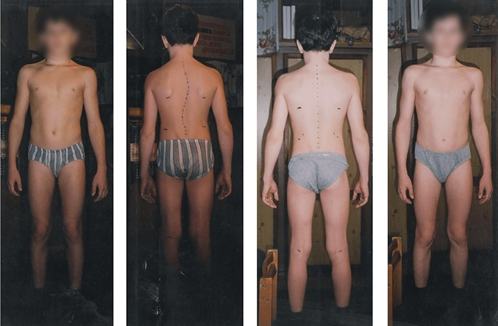
Initial condition shown in the photograph on the left. Four months after completion of treatment, shown on the right. Result: the spinal deformity was eliminated. Posture became correct, the position of the eyes normalized, and the patient reported no complaints.

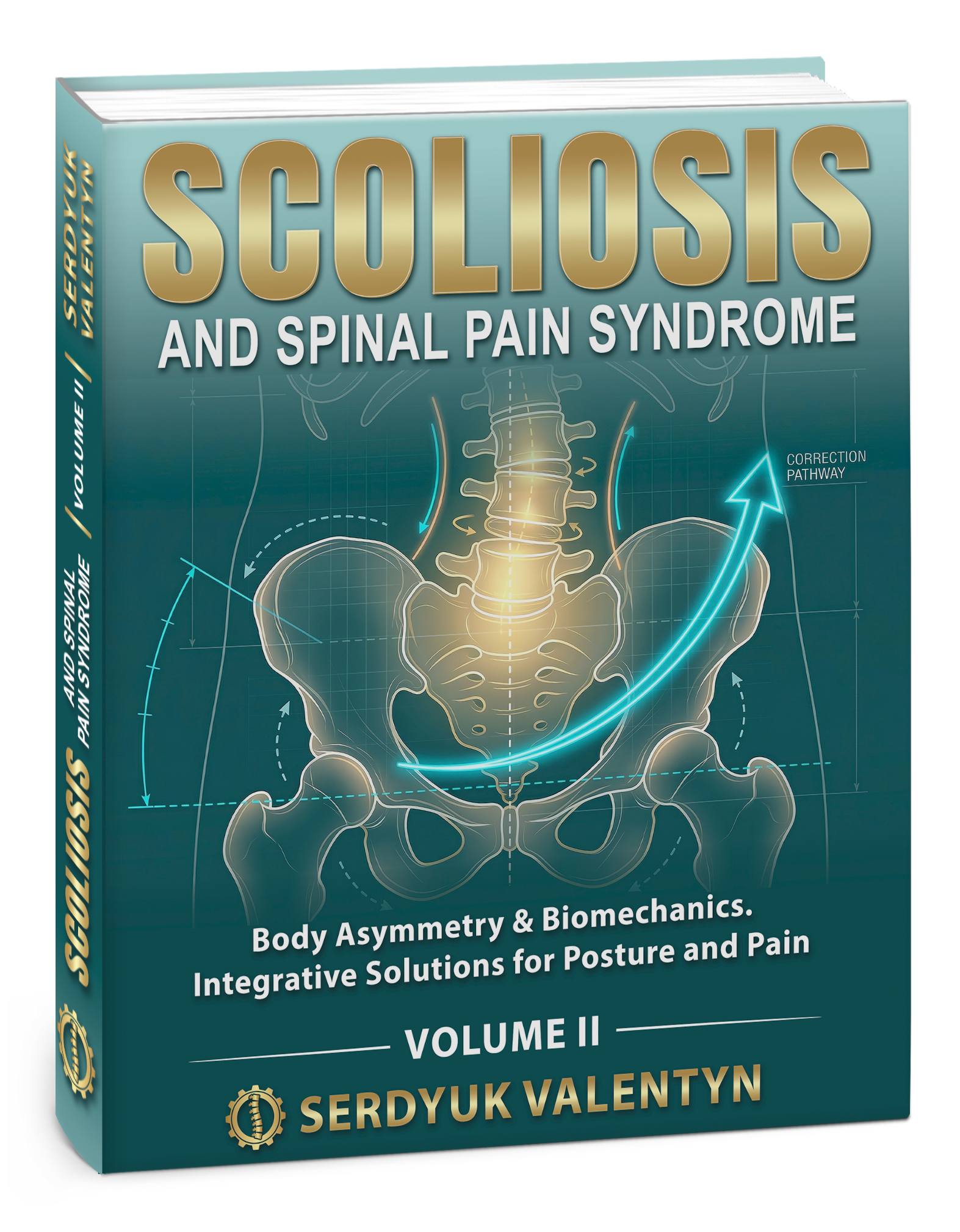
Volumes I & II — Body Asymmetry & Biomechanics, Integrative Solutions for Posture and Pain. For adults, parents, and clinicians seeking a more coherent framework.
Millions of children and adults suffer from idiopathic scoliosis and spinal pain. The author presents two discoveries explaining both scoliosis and many related diseases. Over 25 years, the author treated more than 10,000 patients.
This book is available for purchase on Amazon in both electronic (eBook) and PaperBack formats.

 Buy on Amazon